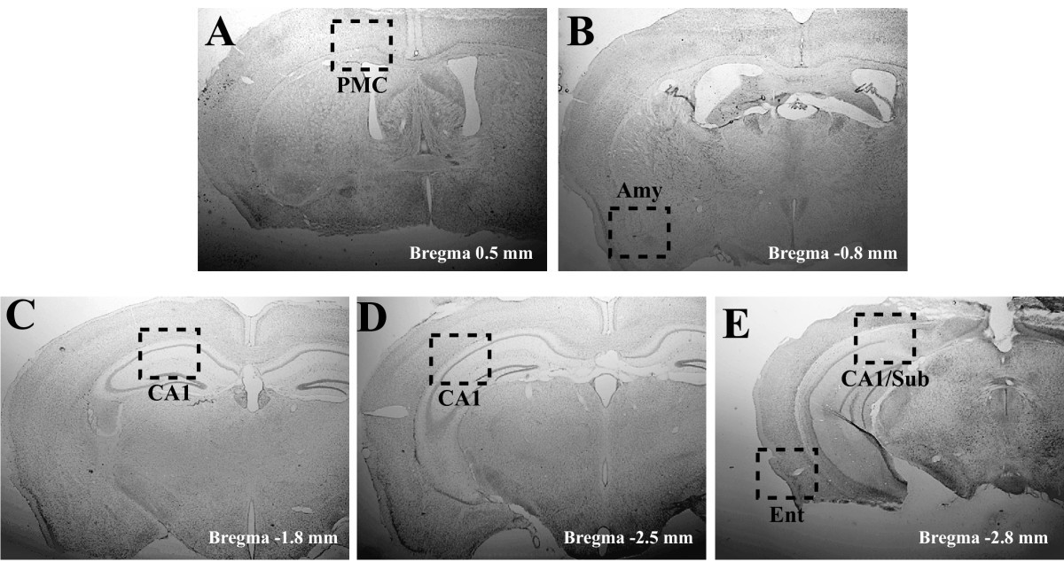
Figure 1

Representative Nissl-stained brain sections from 3xTg-AD mice depicting regions examined by immunohistochemistry in this study. Coronal mouse brain sections (30 μm) were prepared from 3xTg-AD mice sacrificed at 6 months of age and were processed for Nissl staining. Primary motor cortex (PMC) at Bregma 0.5 mm (A), amygdala (Amy) at Bregma -0.8 mm (B), CA1 hippocampal sections at Bregma -1.8 mm (C), at Bregma -2.5 mm (D), and at Bregma -2.8 mm (E), as well as entorhinal cortex (Ent) at Bregma -2.8 mm (E) are outlined by dotted boxes to illustrate the sub-regions of the brains examined in this study. Photomicrographs were obtained at 1.25×.